Dear Dr. Coupe,
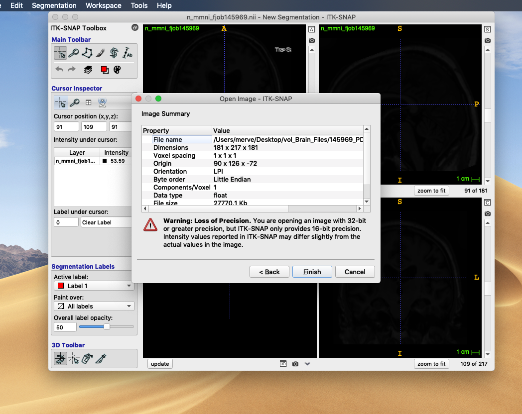
Thank you very much for your response. I run the pipeline for the
sub-optimally registered case, and I used n_mni_fjob file as the
input per your suggestion. It worked well.
I would like to briefly describe how I do the manual corrections
and I will appreciate if you can tell if you find that approach
useful, or have any better ideas. I will also some questions
regarding three cases within our dataset. The questions below will
constitute the backbone of the discussion part of our project, and
since you are the expert on this subject, I wanted you ask for your
opinion.
Manual Correction
Our aim is to understand how shrinked the brain is within the skull
from cross-sectional MRI image. Therefore, extra-ventricular CSF
volume (indicating how sulci are distanced away from each other),
ventricular sizes, and their proportions to cortex GM, cerebral WM
are significant.
On crisp_mni_fjob file, you see some parts of the dura and sagittal
sinus are labeled as GM (green) or CSF (red) (see


). I delete such voxels in all orthogonal planes. Then, I will
check volumes and statistics on ITK-SNAP to see how each volume is
changed. Since, I am only deleting cerebral cortical GM and sulci
CSF, I will subtract/add this difference to the volume we have on
the volBrain report. I think using manually corrected volumetric
result may give a better correlation, if there is. What do you
think about this approach? Is there a path you follow while doing
manual corrections on volBrain files with ITK-SNAP?
Here, this question also emerges: are volumetric results on the
final volBrain report calculated from native crisp or mni
registered crisp file? Since I am doing the manual corrections on
n_mmni crisp file, if the final volumes on the report are
calculated from native space, I need to apply inverse
transformation to the manually corrected mni crisp file and then
add/subtract the volume difference to results on the volBrain
report (or I need to re-do all of the manual corrections on
native_crisp files).
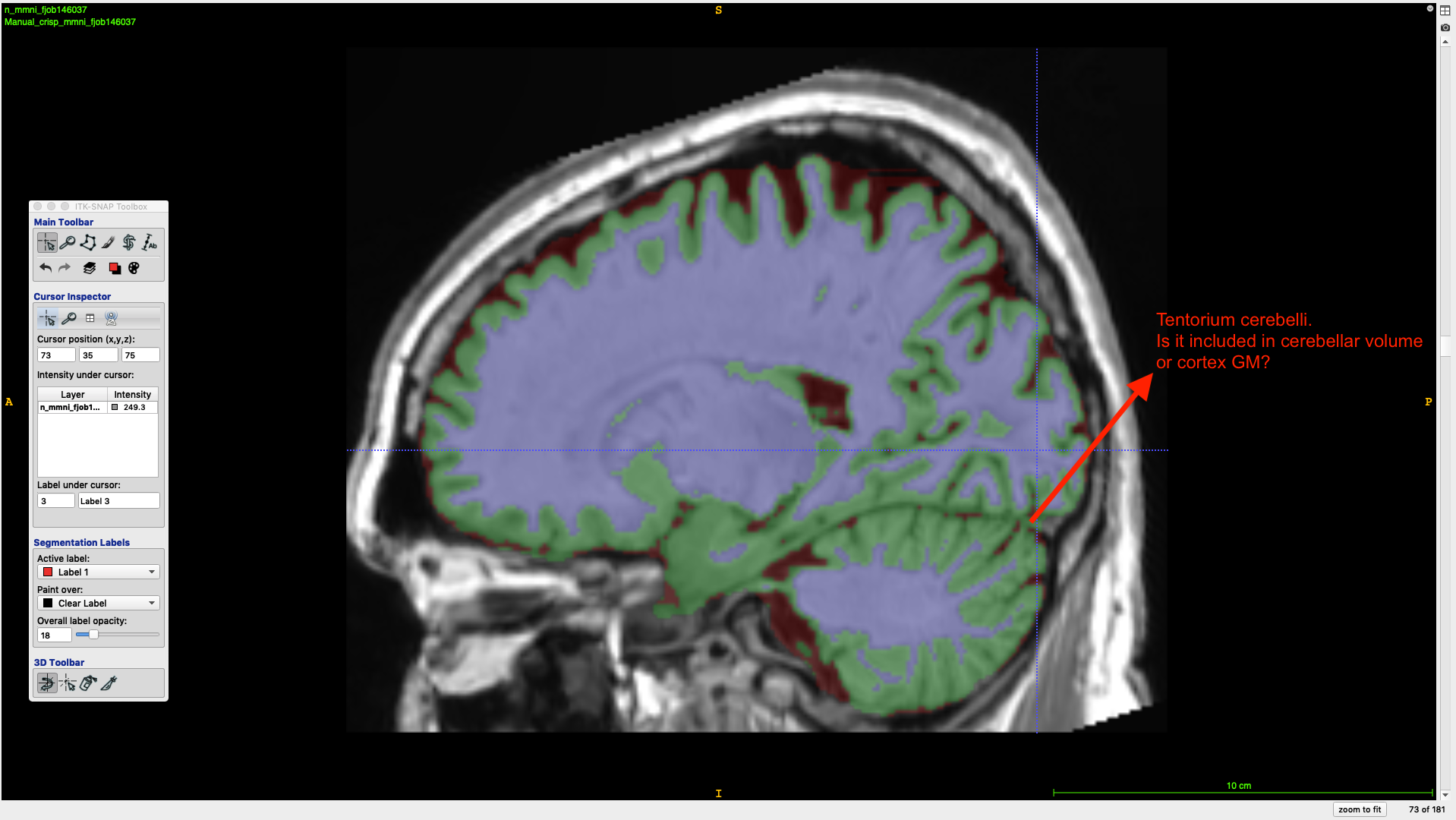
I do not delete the voxels of tentorium cerebelli, since as we
talked, I do not know whether that part is included to the cerebrum
or cerebellum. If it is included to the cerebellum, I should not
subtract it from the GM volume result on volBrain report. But, I am
guessing it is included into the cerebrum.
Questions about 4 Cases
Patient 1 (job146032, Report Date June 10) (Re-do: job151027, Report Date
July 11)
This case was the one that is sub-optimally registered. Using
ITK-SNAP, you can appreciate that the segmentation is affected. I
used n_mni_fjob as input file and it gave a much better
segmentation result and different volumetric results (job151027,
Report Date July 11) (see

).
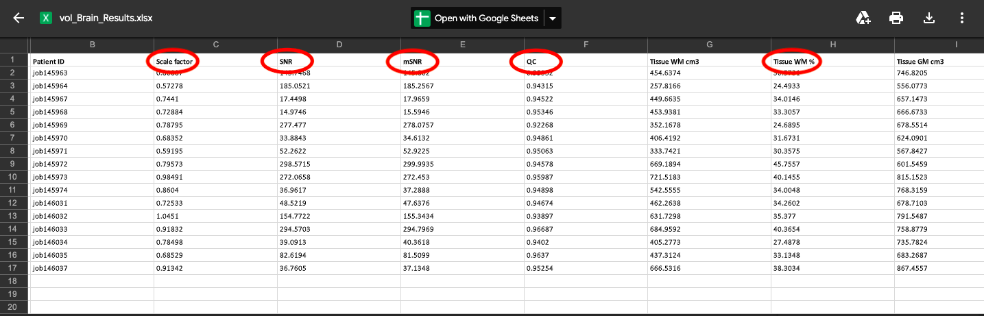
Patient 2 (job145969, Report Date June 9) (Re-do: job151308, Report Date
July 12, Input: n_mmni_fjob)
This case was registered successfully. I wanted to re-do it with
n_mmni_fjob input to see the effect of using normalized, filtered
image as the input in an optimally registered case.
In volBrain results file, the volumes are pretty different, thought
the percentages are almost the same. Is it because the volumetric
calculation is done on the native image? (see

) Since in your paper, the pipeline includes MNI registration
before the tissue segmentation, I am guessing that the segmentation
and volumetric calculations are done on MNI space, then with
inverse transformation, calculated for native space and the final
result on volBrain report is for the native space. Is that correct?
Patient 3 (job145972, Report Date June 9) (Re-do: job151387, Report Date
July 12, Input: n_mmni_fjob)
Though we had no error during the processing, the resulting
segmentation file of this case is problematic. As you can see on

, left frontal lobe is missing and the region supposed to be
labeled as WM is labeled as CSF. It resulted with the similar
problem even after re-running the pipeline with the input n_mmni.
The original image is

. What is your suggestion for this case? Should we do it manually
from scratch?
Patient 4 (job146036, Upload Date June 10) (Re-do: job151028, Upload Date
July 11)
This case could not be segmented at all, and there is no file nor
report available. You can see the error on

. Is there an explanation why this case could not be processed and
the previous one had a problem? Should we do this case manually as
well?
I appreciate any contribution you may have. Thank you very much for
allocation your time.
Best regards,
Merve


